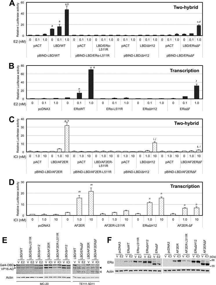
FIGURE 5.
LBD dimer formation and transcription activities. A, HepG2 cells were cotransfected with pG5-luc and expression vector for Gal4-DBD fused WT or mutated ERα LBDs (pBIND-LBD/WT, pBIND-LBD/ERα-L511R, pBIND-LBD/ΔH12, or pBIND-LBD/ERαΔF) in the presence of the expression vector for VP16-AD (pACT) or VP16-AD-fused WT or mutated ERα LBDs (pACT-LBD/WT, pACT-LBD/ERα-L511R, pACT-LBD/ΔH12, or pACT-LBD/ERαΔF). Cells were treated with either vehicle (0 nm) or E2 (0.1 and 1 nm). B, HepG2 cells were cotransfected with 3xERE-TATA-luc, pRL-TK, and expression vector for WT ERα, ERα-L511R, ERαΔH12, or ERαΔF and treated with either vehicle (0 nm) or E2 (0.1 and 1 nm). C, HepG2 cells were cotransfected with pG5-luc and the expression vector for Gal4-DBD-fused AF2ER or mutated ERα LBDs (pBIND-LBD/AF2ER, pBIND-LBD/AF2ER-L511R, pBIND-LBD/ΔH12, or pBIND-LBD/AF2ERΔF) in the presence of the expression vector for VP16-AD (pACT), VP16-AD-fused AF2ER, or mutated ERα LBDs (pACT-LBD/AF2ER, pACT-LBD/AF2ER-L511R, pACT-LBD/ΔH12, or pACT-LBD/AF2ERΔF). Cells were treated with either vehicle (0 nm) or ICI (1 and 10 nm). D, HepG2 cells were cotransfected with 3xERE-TATA-luc, pRL-TK, and expression vector for AF2ER, AF2ER-L511R, ERαΔH12, or AF2ERΔF and treated with either vehicle (0 nm) or ICI (1 and 10 nm). The luciferase activities in A and C are represented as fold change over vehicle in each pACT and pBIND-LBD co-transfected sample. The luciferase activities in B and D are represented as fold change over vehicle in the empty expression vector-transfected (pcDNA3) cells. Luciferase activities are represented as mean ± S.D. a, c, g, i, and k, p < 0.001 against vehicle in each pACT and pBIND-LBD co-transfected sample; b, d, h, j, and l, p < 0.001 against vehicle in each pACT-LBD and pBIND-LBD co-transfected sample; e, f, m, n, and o, p < 0.001 against the vehicle level of each receptor. E, whole cell lysate was prepared from vehicle-treated (V), E2-treated (1 nm), and ICI-treated (10 nm) transfected HepG2 cells and analyzed by immunoblotting with anti-ERα antibody (MC-20 or TE111.5D11) to indicate the expression levels of Gal4-DBD/LBD (closed arrowhead) and VP16-AD/LBD (open arrowhead). β-actin (Actin) was used as a loading control. A representative Western blot analysis is shown. F, whole cell lysates extracted from vehicle-treated (V), E2-treated (1 nm), and ICI-treated (10 nm) transfected HepG2 cells were analyzed by immunoblotting with anti-ERα antibody (H-184, ERα) to indicate the expression levels of ERα WT and mutants. β-actin (Actin) was used as a loading control. A representative Western blot analysis is shown.

