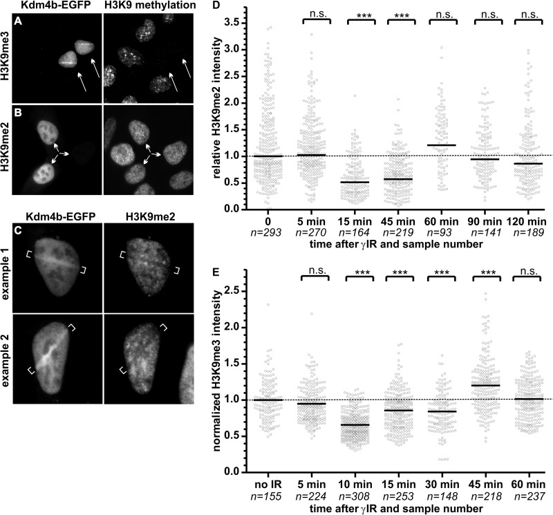
FIGURE 8.
DNA damage-induced changes in H3K9 methylation. 24 h after transfection, U2OS cells expressing low level Kdm4b-EGFP demonstrated a complete loss of H3K9me3 (A) but not H3K9me2 (B). C, U2OS cells were plated on glass-bottom dishes and transfected with Kdm4b-EGFP. 24 h after transfection, DNA damage was induced in EGFP-positive cells using two-photon laser micro-irradiation. Ten minutes after micro-irradiation of the first cell, the plates were fixed and stained for H3K9me2 by immunofluorescence. Two examples are shown. D and E, U2OS plated on glass coverslips were treated with two Gy of γ-irradiation (γIR). Plated cells were fixed at denoted time-points after irradiation. Cells were stained by immunofluorescence for H3K9me3 (D) or H3K9me2 (E), and the nuclei were counterstained with DAPI. H3K9me2 levels were quantified for each nuclei (sample numbers are shown on the graph) using CellProfiler and expressed relative to the mean H3K9me3 levels of the non-irradiated control cells. The mean methylation level was compared with the non-irradiated control using an unpaired t test with Welch's correction for unequal variance. n.s., not significant. p > 0.05. ***, p < 0.0001.

