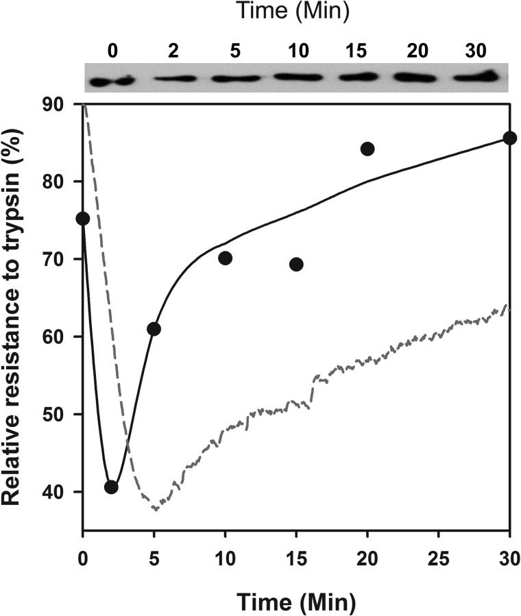
FIGURE 4.
Trypsin treatment of misfolded monomeric luciferase revealed unfolding by Hsp110/Hsp40. Upper panel, Western blots of trypsin-treated luciferase. At the indicated time points of the Hsp110 + Hsp40 + ATP-dependent refolding reaction as in Fig. 2A, the samples were treated for 3 min with 0.04 mg/ml trypsin, separated on SDS gel, and detected on Western blots with luciferase antibodies. Lower panel, the relative semiquantitative luciferase signal at 60 kDa from the Western blot (plain circles). The relative net ThT fluorescence signal of FTluc under the same conditions from Fig. 2B is shown for comparison (red dotted line).

