Figure 4.
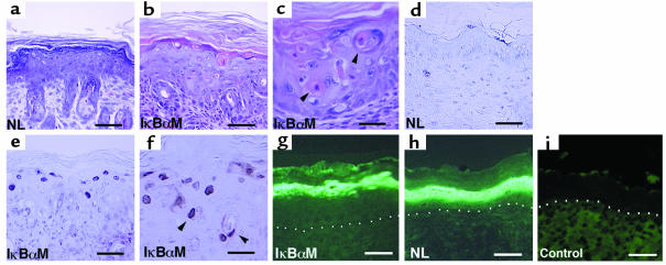
Epidermal apoptosis with blockade of NF-κB function in vivo. (a–c) Histology of skin in vivo. (a) Age- and site-matched control versus (b, c) IκBαM[+] mice transgenic for loss of epidermal NF-κB function. Note the presence of apoptotic cell morphologic changes extending down to the lower spinous layers (arrows) and the complete absence of such changes in the control. (d–f) TUNEL assay in vivo. (d) Age- and site-matched control versus (e, f) mice transgenic for loss of epidermal NF-κB function. Note the presence of TUNEL-positive cells in the granular layer extending down into the lower spinous layers in IκBαM[+] mice. (a, b, d, e) Bars = 50 μm, (c, f) bars = 25 μm. Expression of the terminal differentiation marker loricrin is not inhibited by NF-κB blockade in vivo. Skin from (g) keratin promoter–driven IκBαM[+] transgenic mice (NL) (h) and littermate control were immunostained using antibody specific for loricrin. (i) Anti-mouse secondary antibody alone controls for background immunofluorescence. The dermal-epidermal boundary is highlighted by white dots. (g, h) Bars = 40 μm.

