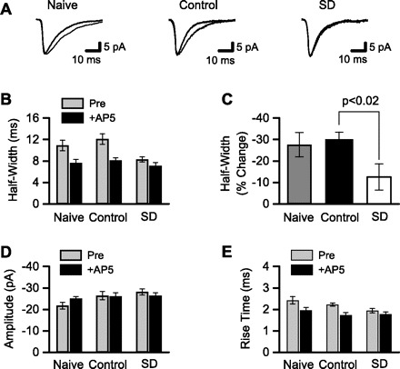
Fig. 2.

NMDAR-mediated, but not α-amino-3-hydroxy-5-methyl-4-isoxazole propionic acid receptor (AMPAR)-mediated, synaptic currents were reduced after sleep deprivation. Whole cell patch-clamp was used to record sEPSCs. A: averaged sEPSCs from 3 different CA1 pyramidal neurons. sEPSCs were recorded in low-Mg2+ ACSF with GABA receptors blocked (thin line) and after addition of the NMDAR antagonist d-AP5 (thick line). Recordings are from single neurons in slices from (left to right): naive animal housed in standard animal cage, control animal kept on large platform over water, and sleep-deprived (SD) animal kept on small platform over water. The d-AP5-sensitive portion of each sEPSC reflects the NMDAR-mediated component of synaptic current. B: NMDAR-mediated synaptic currents were quantified by measuring EPSC half-width before and after application of d-AP5. sEPSC half-widths were averaged across all cells from the same treatment condition. d-AP5 substantially reduced sEPSC half-width in cells (n = 11, 9) from naive and control animals, but not in cells (n = 9) from SD animals. C: mean change in sEPSC half-width after d-AP5 application was calculated from the data shown in B. The change in half-width was significantly smaller in the SD group compared with control. sEPSC amplitudes (D) and rise times (E) are shown. After NMDARs were blocked by d-AP5, there were no differences among the 3 groups in sEPSC half-width (B), amplitude (D), or rise time (E), indicating that there was no effect of treatment on AMPAR function.
