
Fig. 3.
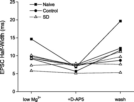
In cells that could be held long enough for complete washout of d-AP5, NMDAR function recovered to baseline levels. sEPSC half-width is plotted for each cell that was held long enough to complete washout of d-AP5. In cells from naive and control animals, sEPSC half-widths were consistently and substantially reduced from baseline (low Mg2+) during d-AP5 application as shown earlier (see Fig. 2), but fully recovered following washout of d-AP5 (wash). Even in this reduced sample size, it is evident that sEPSC half-widths in cells from SD animals were less sensitive to d-AP5 application, as shown above in Fig. 2; however, when half-width was reduced by d-AP5 in these cells, it recovered completely after washout.