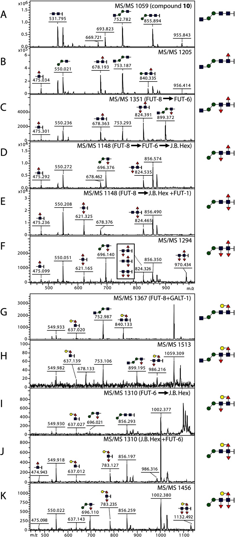
FIGURE 8.
MS/MS of substrates and products elucidating the formation of trifucosylated glycans. Spectra A–F display the fragmentation patterns of the non-galactosylated glycans Hex2HexNAc2–3Fuc0–3-(CH2)5NH2 with m/z 1059, 1205, 1351, 1148, and 1294 (see Fig. 7, A and B), whereas spectra G–K display those of the galactosylated glycans Hex3HexNAc2–3Fuc0–3-(CH2)5NH2 with m/z 1367, 1513, 1310, and 1456 (see Fig. 7, C and D). All the ions annotated are [M + Na]+, and predicted structures of the key ions are shown in Consortium for Functional Glycomics format; the alkylamine linker, -(CH2)5NH2, is represented by a short vertical bar at the right of N-acetylglucosamine. 475, HexNAc1Fuc1-(CH2)5NH2; 621, HexNAc1Fuc2-(CH2)5NH2; 637, Hex1HexNAc1Fuc1-(CH2)5NH2; 678, HexNAc2Fuc1-(CH2)5NH2; 696, Hex2HexNAc1Fuc1; 783, Hex1HexNAc1Fuc2-(CH2)5NH2; 824, HexNAc2Fuc2-(CH2)5NH2; 899, Hex2HexNAc2Fuc1; 970, HexNAc2Fuc3-(CH2)5NH2; 1132, Hex1HexNAc2Fuc3-(CH2)5NH2. Red triangles, fucose; yellow circles, galactose; blue squares, N-acetylglucosamine; green circles, mannose.

