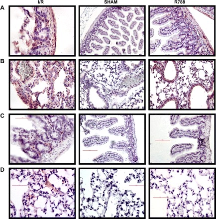
Fig. 3.

Syk staining in intestinal and lung tissues. Tissue sections were stained as described in materials and methods. A and B: Syk staining was assessed in intestinal (A) and lung (B) tissues by immunohistochemistry staining. Magnification: ×400 for I/R and R788 in A, ×200 for Sham in A and all of B. C and D: phosphorylated (p-)Syk staining was performed in intestinal (C) and lung (D) tissues by immunohistochemistry. All photomicrographs are ×200 magnification.
