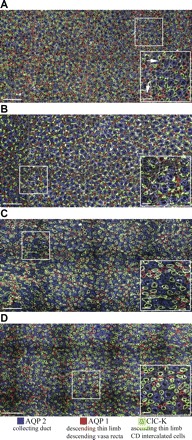
Fig. 1.

Immunolocalization of loops of Henle and collecting ducts (CDs) in inner medullary transverse sections. A and B: kangaroo rat, 500 and 2,000 μm below the outer medulla (K rat 2 in Fig. 4). C and D: Munich-Wistar rat, 500 and 2,000 μm below the outer medulla (MW rat 5 in Fig. 4). Antibodies were applied at equal concentrations, and images were acquired at equivalent intensity scaling. Structures not labeled with aquaporin 1 (AQP1), aquaporin 2 (AQP2), or the chloride channel ClC-K antibodies are shown in off-white, labeled with wheat germ agglutinin. Scale bars: 100 μm and 20 μm (inset). Boxed areas are enlarged in right corner insets. Arrows identify AQP1-positive descending vasa recta (DVR).
