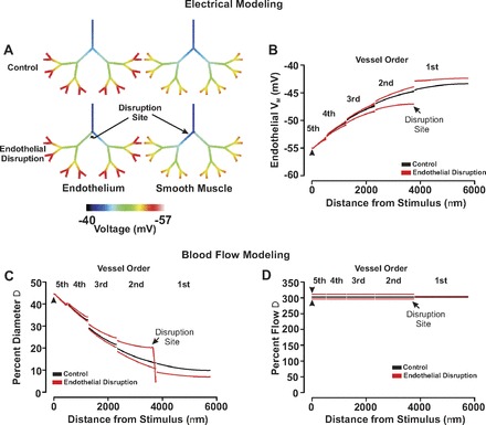
Fig. 11.

The effect of focal endothelial disruption on the ascension of responses in a virtual arterial network. A and B: for simulations, an endothelial segment in all of fifth-order arteries was voltage clamped 15 mV negative to resting Vm (−40 mV, 200 ms) under control conditions and following endothelial ablation. Endothelial (solid line) and smooth muscle (dotted line) voltage responses were displayed in steady state with the use of color maps (A) or 2-dimensional plots (B). C and D: for simulations, vasomotor and blood flow responses were calculated for the virtual network under conditions in which all fifth-order arteries were voltage clamped 15 mV negative to the resting Vm (−40 mV, 200 ms). The endothelial layer was intact or focally disrupted at a site distal to the first- and second-order branch. Flow data are presented in absolute terms (C) or as a percent change from baseline (D). Arrow heads denote site of stimulation.
