Fig. 2.

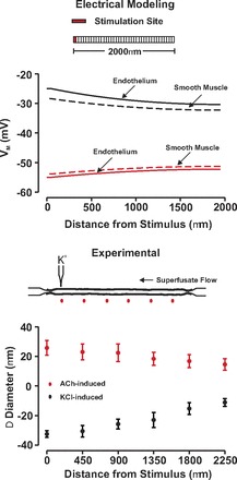
Endothelial-initiated responses conduct along resistance arteries independently of the stimuli polarity. Top: for simulations, endothelial cells within 1 arterial segment were voltage clamped 15 mV negative (red) or positive (black) to resting Vm (−40 mV, 200 ms) while steady-state endothelial (solid line) and smooth muscle (dotted line) electrical response was monitored along the vessel. Bottom: for experimentation, ACh (red, 10 mM) or KCl (black, 250 mM) was pressure ejected onto a small region of a fifth-order mesenteric artery. Vasomotor responses were monitored at sites 0, 450, 900, 1,350, 1,800, and 2,250 μm distal to the point of agent application. Resting, minimal, and maximal diameters were as follows ACh (n = 4): 108 ± 5 μm; 18 ± 2 μm; 140 ± 7 μm; KCl (n = 6): 108 ± 5 μm; 17 ± 1 μm; 138 ± 7 μm.
