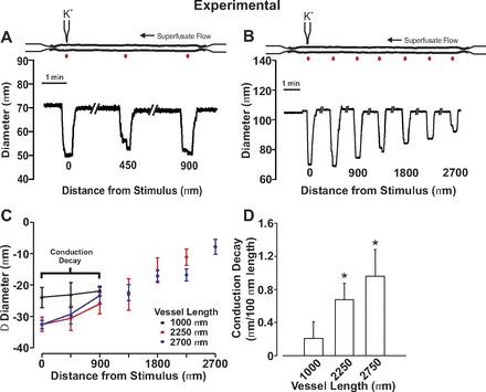
Fig. 5.

Electrical decay increases with the length of mesenteric arteries. For experimentation, endothelial cells in a small portion of a fifth-order mesenteric artery were focally stimulated with KCl while vasomotor responses were monitored along the artery. A and B: representative traces showing conducted constriction in arteries of different length. C and D: summary data were plotted as a change in vessel diameter (C) or as conduction decay (D). Resting, minimal, and maximal diameters were as follows: 1,000 μm in length (n = 7), 101 ± 7 μm, 24 ± 4 μm, 134 ± 7 μm; 2,250 μm in length (n = 6), 109 ± 7 μm, 27 ± 4 μm, 134 ± 8 μm; 2,700 μm in length (n = 6), 114 ± 8 μm, 32 ± 6 μm, 140 ± 9 μm. *Significant difference from 1,000 μm.
