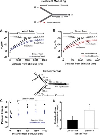
Fig. 6.

Electrical decay in a branching arterial structure. A and B: for simulations, an endothelial segment in 1 or both daughter arteries was voltage clamped 15 mV positive to resting Vm (−40 mV, 200 ms). Steady-state depolarization was plotted along the branching structure and compared with a similar length of unbranched virtual artery. C and D: for experimentation, 1 fifth-order mesenteric artery from a fourth- to fifth-order branch point was stimulated with KCl (250 mM, 20-s pulse) while vasomotor responses were monitored along the network. Summary data were plotted as a percent change of vasoconstrictor capacity (C) or as conduction decay (D). Resting, minimal, and maximal diameters for the branching arterial structure (n = 9) were as follows: parent vessel, 152 ± 10 μm, 56 ± 7 μm, 207 ± 10 μm; stimulated fifth-order artery, 105 ± 9 μm, 36 ± 8 μm, 160 ± 14; unstimulated fifth-order artery, 114 ± 10 μm, 33 ± 6 μm, 147 ± 9 μm. For the unbranched fifth-order vessels (n = 7), the resting, minimum, and maximal diameters were 121 ± 10 μm, 32 ± 6 μm, and 161 ± 15 μm, respectively. *Significant difference from the unbranched artery.
