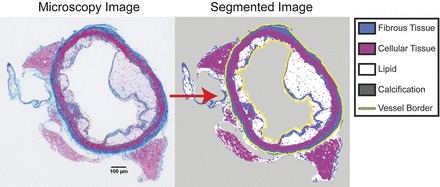
Fig. 1.

Semi-automated segmentation of atherosclerotic lesions: a sample cross-section of an atherosclerotic low-density lipoprotein receptor-deficient (LDLR−/−) mouse aorta stained with a combination of Masson's trichrome and Von Kossa's calcium stain. The microscopy image (left) was loaded into custom Matlab software and segmented (right) using a semi-automated algorithm. In brief, tissue types were automatically identified using a k-means clustering algorithm and then touched up (for example, to distinguish lipid from background) using an implementation of the Live Wire algorithm (7). The perimeter of the lumen and adventitia were also identified with Live Wire (yellow line).
