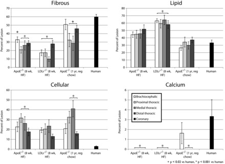
Fig. 4.

Relative composition of atherosclerotic plaques in mice and humans. Based upon segmented atherosclerotic plaques (Fig. 1), we calculated the relative composition of lesions [fibrous (top left), lipid (top right), cellular (bottom left), and calcified (bottom right)] for humans and mice. Error bars represent SE. Tukey's honestly significant difference test results are noted for comparisons of brachiocephalic aorta vs. human coronary and all descending aortae pooled together vs. human coronary.
