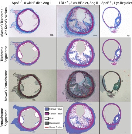
Fig. 7.

Validation of staining protocols. We stained consecutive serial sections of mouse tissue with Masson's trichrome and Von Kossa's calcium protocols (top) and with Movat's pentachrome protocol (bottom). Representative images paired with their segmentation demonstrated qualitative agreement. The location of local maxima of stress (not shown) did not differ between specimens. Only tissue inside the lines marked “vessel border” was included in our study, so periadventitial segmentation differences will not affect results. A representative problem with the Movat's pentachrome stain in our tissues is visible in the LDLR−/− and older ApoE−/− specimens: the vascular casting compound absorbed dye, partially obscuring nearby tissue.
