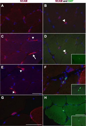
Fig. 3.

Neural cell adhesion molecule (NCAM) and Ki67 double stainings. The immunohistochemical (IHC) staining of 7-μm cross sections of muscle biopsies with the use of antibodies against NCAM (red), Ki67 (green), and 4′,6-diamidino-2-phenylindole (DAPI; blue) is shown. The left images are examples of diverse NCAM staining of (mostly) satellite cells. A and C: examples of NCAM+ satellite cells, with the typical appearance of a blue nucleus surrounded by red NCAM staining. Some satellite cells show extensions around the muscle fiber periphery (arrowhead). C: nerve staining by the NCAM antibody (arrow). E: three NCAM+ satellite cells, two with cytoplasmic extensions (arrowheads). G: an NCAM+ satellite cell and a myofiber with NCAM+ staining of the membrane and cytoplasm. The right images show combinations of NCAM (red), Ki67 (green), and DAPI (blue) staining. B and D: double staining of the same section showing, in B, two satellite cells (NCAM+, red); one is also a Ki67+ cell (green), as in the merged image in D (arrowhead). D, inset: Ki67 staining of that proliferating cell. F: two proliferating cells in the extracellular matrix. F, inset: Ki67 staining alone. H: Ki67+ cell located in the extracellular matrix. H, inset: Ki67 staining alone. Scale bars = 50 μm.
