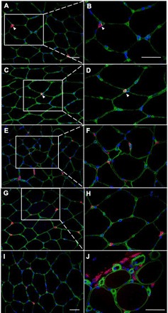
Fig. 4.

CD16 (neutrophils) and CD68 (macrophages) double stainings with laminin. IHC staining of 7-μm cross sections of muscle biopsies with the use of antibodies against CD16 (A–F) or CD68 (G–J) (red), laminin (green), and DAPI (blue) is shown. The left images are at low (×20) magnification, and the right images are at higher (×40) magnification. Part of the section on the left is enlarged on the right where indicated. A–F: CD16 staining (red). A and B: sections from a preexercise biopsy showing one CD16+ cell (arrowhead) outside the basal lamina (laminin, green) of muscle fibers. C and D: sections from a day 8 biopsy (NSAID leg) showing one CD16+ cell (arrowhead). E and F: sections from a day 8 biopsy (NSAID leg) with many CD16+ cells. All are located outside the basal lamina. Cells were counted as CD16+ cells only when they also had a visible nucleus (DAPI, blue). CD16+ cells were never observed inside muscle fibers. G–J: CD68 staining (red). G and H: sections from a preexercise biopsy showing examples of CD68+ cells. All are located outside the basal lamina (green). I: section from day 8 biopsy (no block leg) showing some CD68+ cells. J: section from day 8 biopsy (NSAID leg) showing several CD68+ cells as an example of the most extreme amount of inflammatory cells observed. CD68+ cells were never observed inside muscle fibers. Scale bars = 50 μm.
