
Fig. 1.
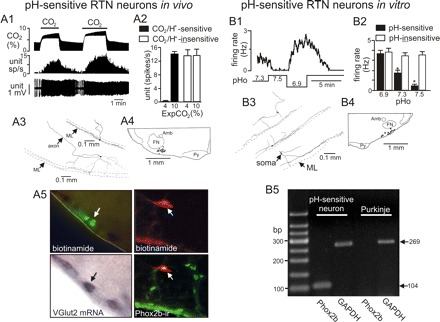
Defining characteristics of RTN chemoreceptors in vivo and in vitro. A1: the typical firing rate response of a CO2/H+-sensitive neuron in vivo to changes in end expiratory CO2; increasing Exp CO2 increased neuronal activity in a reversible and repeatable manner. A2: average firing rate of CO2/H+-sensitive (n = 26) and CO2/H+-insensitive (n = 39) neurons in vivo under control (4% CO2) and hypercapnic (10% CO2) conditions. After recording cells were labeled with biotinamide for later conformation of location, morphology, and neurochemical phenotype. A3: the structure of two CO2/H+-sensitive neurons labeled in vivo (ML, marginal layer). A4: plots the location of 17 CO2/H+-sensitive neurons (black dots) recorded in vivo. Bregma level −11.4 mm (Amb, nucleus ambiguous; FN, facial nucleus; ML, marginal layer; Py, pyramidal tracts). A5: a biotinamide (Alexa Fluor 488 fluorescence)-labeled CO2/H+-sensitive RTN neuron recorded in vivo (top) and the same cell expresses vesicular glutamate transporter-2 (VGLUT2) mRNA (bright-field illumination; bottom). A6: top shows a CO2/H+-sensitive RTN neuron recorded in vivo and labeled with biotinamide (Cy-3, red), and bottom shows that the same cell is immunoreactive for Phox2b (Alexa 488, green), biotinamide with Cy-3 (red); colocalization is shown in yellow. B1: trace of firing rate and bath pH show characteristic responses of a pH-sensitive RTN neuron to pH values ranging from 6.9 to 7.5; these cells are spontaneously active at control pH 7.3, nearly silent at pH 7.5, and robustly active at pH 6.9. B2: average firing rate of pH-sensitive (n = 40) and pH-insensitive (n = 47) neurons recorded in vitro at varying pH. *P <0.01 for effect of pH. B3: structure of three biocytin-filled pH-sensitive neurons that are reminiscent of CO2/H+-sensitive RTN neurons recorded in vivo. B4: composite map shows the location of 11 pH-sensitive neurons (black dots) recorded in vitro (Amb, nucleus ambiguous; FN, facial nucleus; ML, marginal layer; Py, pyramidal tracts). B5: agarose gel of single cell RT-PCR for Phox2b and GAPDH; the chemosensitive RTN neuron expresses Phox2b, but the Purkinje cell does not. This figure is composed of previously published data (38, 39, 59) and presented here with permission from the appropriate journals.