
Fig. 5.
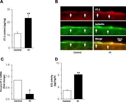
Effect of IH on endothelin-1 (ET-1) expression in the carotid sinus region. A: average data of ET-1 content measured by enzyme immunoassay (EIA) in the carotid sinus region from control and IH-exposed rats. B: example illustrating the immunocytochemical localization of ET-1-like immunoreactivity and isolectin labeling of endothelial cells in the carotid sinus region (at arrows) from control (left) and IH-treated (right) rat. C: effect of IH on pre-pro ET-1 mRNA levels in the carotid sinus region. D: IH increased endothelin converting enzyme (ECE) activity in the carotid sinus region. Data are means ± SE from control and IH rats; n = 6 rats in each group. Significant difference compared with normoxia-treated rats (control): *P < 0.05; **P < 0.01.