
Fig. 2.
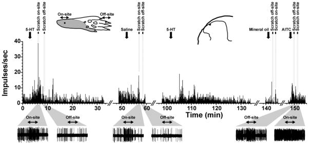
Individual example. Upper: peristimulus-time histogram (PSTHs; bin width: 1 sec) of unit located in superficial dorsal horn. Downward arrows indicate time of application of indicated stimuli. Times of on- and off-site scratching are indicated by black bars with dashed lines. Upper left inset: drawing of hindpaw showing receptive field (gray) and locations of on-site and off-site scratching (dots and horozintal two-headed arrows). Upper right inset: recording site (dot) on drawing of lumbar spinal section. Lower: Gray trapezoids beneath PSTH indicate 60-sec time windows that have been expanded to show spike traces (bottom row) of neuron’s response before, during and after scratching. Scratch period indicated by two-headed arrows.