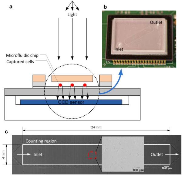
Fig. 2.
A schematic view of the CCD imaging platform: (a) CCD imaging platform to detect the captured cells. When light is incident on the captured cells, the cell membrane diffracts and transmits light. A shadow of the captured CD4+ T-lymphocytes generated by diffraction can be imaged by the CCD in one second. Image is obtained with the lens-less CCD imaging platform. (b) Picture of microfluidic chip and CCD imaging platform. Field of view of the CCD sensor is 35 mm × 25 mm. The entire microfluidic device can be imaged without alignment by simply placing the microfluidic channel on the sensor. (c) Image taken with the lens-less CCD imaging platform and magnified view at the microfluidic channel centre is shown. The magnified picture shows an image obtained by diffraction. Scale bar, 100 μm.

