
Fig. 5.
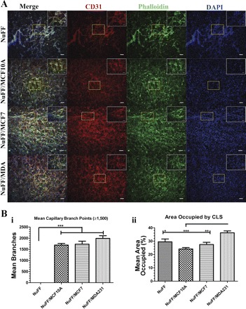
Vascular morphogenesis on ECM deposited by NuFF and cocultures of NuFF with BCCs. Capillary-like structures (CLSs) formed on deposited ECM were visualized with immunostaining for phalloidin (green), CD31 (PECAM; red), and nuclei (DAPI; blue) (A) and quantified for mean capillary branches (≥1,500; i) and % area occupied by capillaries (ii) (B). Scale bars are 100 μm and 50 μm for image insets. Significance levels were set at *P < 0.05, **P < 0.01, and ***P < 0.001. Values shown are means ± SE.