
Fig. 6.
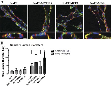
Characterization of capillary lumens from vascular structures. Tubular CLS formed on deposited ECM. A: 3-dimensional z-stack of confocal images (phalloidin in green, CD31 in red, and nuclei in blue) revealed lumens in CLSs grown on all tested ECM as demonstrated by the cross section image of the circled area. Scale bars are 10 μm for cross section images. B: average lumen diameter for the long axis of the tubes was significantly larger for CLSs grown on MDA231-NuFF-derived ECM. Significance levels were set at *P < 0.05. Values shown are means ± SE.