
Fig. 8.
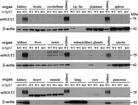
Absence of SGLT2 protein expression in extrarenal organs of the mouse. Western blot analysis was performed in protein lysate of various organs from 2 male and 2 female WT mice. Kidneys of WT mice served as positive control, and kidneys of KO (Sglt2−/−) mice were used as negative control. In the kidneys of WT mice the rSGLT2-Ab recognized a protein band of the predicted size of SGLT2 (∼75 kDa), whereas in other organs the antibody recognized additional unspecific bands. In none of the studied extrarenal WT organs was a band consistently detected that corresponded in size to the SGLT2-related band detected in WT kidney. Each lane was loaded with 10 μg of whole organ lysate. β-Actin was used to confirm equal loading, although its expression varies between organs. i.p., Intraperitoneal.