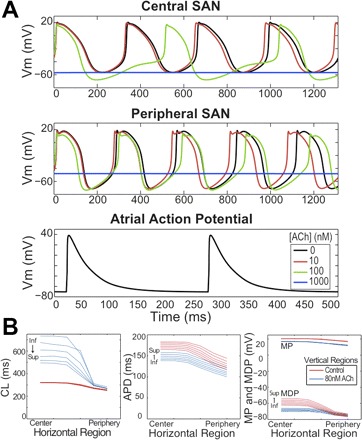
Fig. 2.

SAN and RA isolated cell action potentials. A: top to bottom: intrinsic action potentials (APs) from the central and peripheral SAN and RA free wall under various ACh concentrations ([ACh]). Note that [ACh] was not introduced into the free wall. B: intrinsic SAN AP properties under 0 (red) and 80 (blue) nM [ACh]. From left to right, cycle length (CL), action potential duration (APD), minimum diastolic potential (MDP), and maximum potential (MP) as functions of position within the SAN. Labels on each group of curves indicate inferior (Inf.) and superior (Sup.) regions. For orientation purposes, the crista terminalis would run along the right side of the SAN from superior vena cava to inferior vena cava. Vm, transmembrane voltage.
