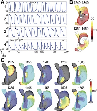
Fig. 5.

Counterclockwise reentry after vagal stimulation. A: voltage traces were obtained at locations labeled in B upper. An atrial premature beat (APB) occurs leading to conduction block labeled in A1. Arrows indicate direction of propagation. Cycle lengths are indicated in A4 to show the initiation and termination of the reentry. B: activation maps for 2 cycles of reentry over the periods (ms) indicated. C: voltage maps taken at the times indicated in ms since ACh application. 10-mV isopotentials are drawn.
