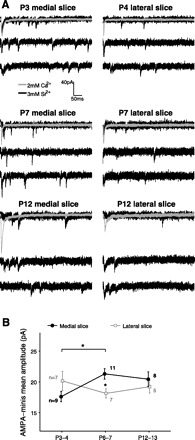
Fig. 2.

Developmental profiles of evoked AMPA miniature currents (“AMPA-minis”) in the medial and lateral SC. A: examples of evoked AMPA-minis recorded from medial (left) and lateral (right) slices at different ages. In these experiments, we first detected a stable evoked AMPA response using a Ca2+-ACSF bath (gray, average trace over 10–20 sweeps). We then replaced extracellular Ca2+ by Sr2+ to desynchronize synaptic release and recorded “miniature,” or evoked quantal AMPA currents (black). B: AMPA-mini amplitudes varied with age in medial but not lateral SC [medial slice: F(2, 25) = 4.18, P = 0.027, lateral slice: F(2, 16) = 0.76, P = 0.48; 1-way ANOVA]. Specifically, AMPA-mini amplitudes increased from P3–4 to P6–7 in the medial but not the lateral slice (P = 0.025, P = 0.45 for medial/lateral slices, respectively, Tukey's post hoc test). At P6–7, AMPA minis were significantly larger in the medial than in the lateral slice (P = 0.021; 2-tailed Student's t-test with false discovery rate procedure for multiple comparisons). Number of cells per group is indicated. Only 1 cell per slice and 1 slice per animal were used. *P < 0.05.
