
Fig. 3.
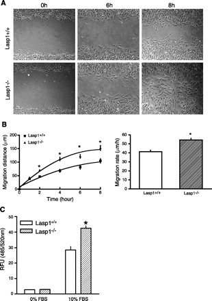
Migration rates of embryonic fibroblasts isolated from wild-type and Lasp1-null littermates assessed using monolayer wound healing and Transwell cell migration assays. A: monolayer wound healing assay: mouse embryonic fibroblasts (MEFs) were plated in 35 mm culture dishes and grown to confluence. A “wound” was generated in the center of each dish with a Gilson P-200 pipette tip, and migratory progress was recorded by phase-contrast microscopy. Representative images from selected time points postwounding are shown in the figure. B: quantitation of data from wound healing assays. Left: time course of MEF migration. Right: rate of migration (changes in the width of the wound over time). There was a significant difference in the distances covered by Lasp1−/− MEFs compared with Lasp1+/+ MEFs. *P < 0.01; n = 4. C: modified Boyden chamber assays: Innocyte Cell Migration chambers were used to measure cell migration over a 6 h period as described in material and methods. Lower chambers containing serum-free media were used as negative controls. The number of MEFs that migrated through the membranes of the chamber inserts during this time period was significantly greater for Lasp1−/− MEFs compared with Lasp1+/+ MEFs. *P < 0.05; n = 3.