
Fig. 4.
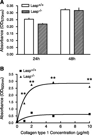
Cell proliferation and adhesion in Lasp1+/+ and Lasp1−/− MEFs. A: cell proliferation was analyzed using a Vybrant MTT Cell Proliferation Assay (Invitrogen). There was no significant difference in proliferation between Lasp1+/+ and Lasp1−/− MEFs at either 24 or 48 h after cell plating. P > 0.05; n = 4. B: the attachment of both Lasp1+/+ and wild-type MEFs was positively correlated with increases in collagen type I concentrations; however, the attachment of Lasp1−/− MEFs was significantly greater than Lasp1+/+ MEFs at all collagen concentrations tested. **P < 0.01; n = 4.