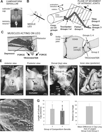
Fig. 1.

Structure of leg and trochanteral campaniform sensilla. A: campaniform sensilla monitor strains in the exoskeleton through the dendritic insertion of the sensory neuron to a cuticular cap. B: drawing of middle leg and locations of campaniform sensilla (redrawn after Cruse and Bartling 1995). The leg is segmented and attached to the body at the basal segment (coxa). The body-coxa joint has considerable freedom of movement. The more distal leg articulations are intrinsic hinge joints that act in a single plane. The distribution of groups of campaniform sensilla is not uniform. No groups are found on the coxa, many groups (Groups 1–4) are concentrated on the trochanter, and single groups are found on the distal segments or subsegments (Groups 5–11). CS, campaniform sensilla. C: interior view of the thorax and muscles that act on the leg (redrawn after Marquardt 1940). The largest muscles that move the leg (retractor, protractor, levator, and depressor muscles) are located in the thorax and insert onto the coxa (Cx) and trochanter (Tr). D: drawing of anterior side of the coxa and trochanter. The groups of campaniform sensilla are located distal to the coxo-trochanteral joint condyle and are therefore in the leg plane. E: reconstructions of confocal images of the exoskeleton. i: Anterior view of trochanter—Group 2 sensilla are located on the anterior side of the trochanter opposite the anterior condyle of the coxo-trochanteral hinge joint. ii: Posterior view—Group 1 receptors are situated on the posterior side and have a similar orientation relative to the posterior joint condyle. iii: Dorsal view—Groups 3 and 4 are located on the dorsal side of the trochanter adjacent to the joint with the femur. iv: Interior view of the posterior half of the trochanter—a thick internal projection (buttress) reinforces the trochanter adjacent to the insertion of the trochanteral depressor muscle. F: scanning electron micrograph showing cuticular caps of Groups 3 and 4. G: histograms of sizes and orientation of sensilla. Each group contains cuticular caps of different sizes (left), and the largest caps of Groups 3 and 4 are mutually perpendicular (right) [mean difference between Groups 3 and 4 = 93.8 ± 13.4° (SD), N = 8].
