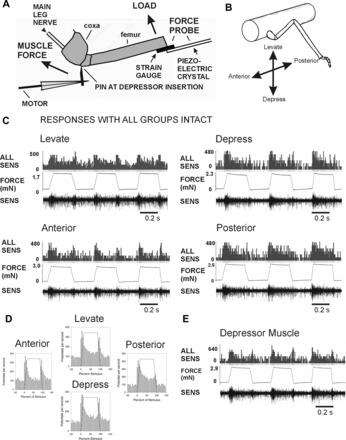
Fig. 2.

Preparation and responses of sensilla with all groups intact. A: preparation. Loads were applied to the femur with a force probe linked to a piezoelectric crystal. The probe contained strain gauges and was used to monitor forces in all experiments. Movement of the leg was resisted by a pin inserted into a small hole adjacent to the depressor muscle insertion. In experiments emulating the effects of muscle contractions, forces were applied to the pin via a computer-controlled linear motor. Sensory activities were recorded in the main leg nerve (nervus cruris) proximal to the coxa; the nerve was crushed close to the mesothoracic ganglion. B: orientation of forces relative to the leg plane. Loads were applied in different directions (levation, depression, anterior, posterior) by rotating the micromanipulator that held the force probe. C: recordings during load application with all groups of campaniform sensilla intact (other receptors ablated). Applying loads via the probe with ramp and hold functions produced sensory discharges in all directions. D: mean sensory discharges: histograms plotting the mean sensory discharges during application of forces in each direction. Discharges occurred during the rising phase that rapidly adapted to lower levels during the hold phase. Prominent phasic discharges occurred to decreasing forces in all directions. E: response to forces applied at the depressor muscle insertion. Intense firing at similar amplitudes occurred when movements of the femur were blocked by the force probe. Bursts were also present during force decreases.
