Fig. 1.

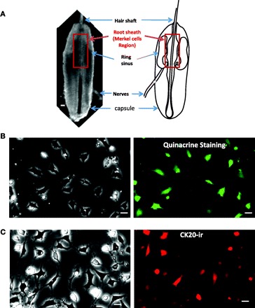
Quinacrine staining and cytokeratin 20 (CK20) immunoreactivity (ir) of cultured Merkel cells dissociated from vibrissal hair follicles of rat whisker pads. A, left: an intact vibrissal hair follicle dissected out from a rat whisker pad and viewed under a light microscope. Right: schematic diagram illustrates the structures of a vibrissal hair follicle. Highlighted region in both panels is the root sheath of upper hair follicle segment where Merkel cells are present at a high density. B: cultured cells labeled with quinacrine. Left: viewed under a light microscope. Right: viewed under a fluorescent microscope. C: cells after immunostaining with an antibody against CK20. Left: viewed under a light microscope. Right: viewed under a fluorescent microscope. In both B and C cells were in culture for 48 h before staining. Scale bars, 50 μm in A and 10 μm in B and C.
