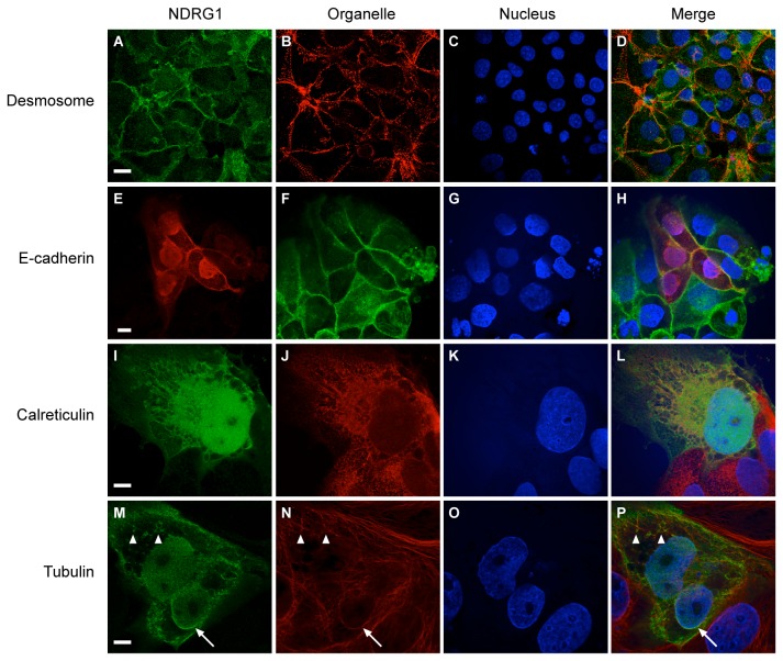
Figure 4. The expression of NDRG1 in cytoplasmic membranes or membrane-associated structures in hypoxic (24 h) trophoblastic lines.
The nuclei (blue), in all panels, were detected using Hoechst 33342 and each organelle was detected using a specific antibody as described in Materials and Methods. (A-D) NDRG1 (green) co-localizes with desmosomes (red) in JEG-3 cells. (E-H) Myc-tagged NDRG1 (red) co-localizes with E-Cadherin (green) in BeWo cells. (I-L) Myc-tagged NDRG1 (green) co-localizes with calreticulin (red) in BeWo cells. (M-P) Myc-tagged NDRG1 (green) co-localizes with tubulin (red) in BeWo cells. Arrow points to perinuclear NDRG1 and tubulin signals. Arrowheads point to peripheral microtubules. Data are representative of at least three independent experiments. Bar = 20 µm in panels A-H, and Bar = 10 µm in panels I-P.

