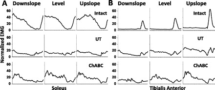
Fig. 3.

Muscle activity during walking at different slopes before and after injury. Normalized Sol (A) and tibialis anterior (TA; B) EMG activity is shown from Intact rats (n = 8), and postinjury Untreated (UT) rats (n = 3) and ChABC-treated rats (n = 4) 10 wk after sciatic lesion. Tracings start and end with paw contact and have been time-normalized to 100 x-axis units.
