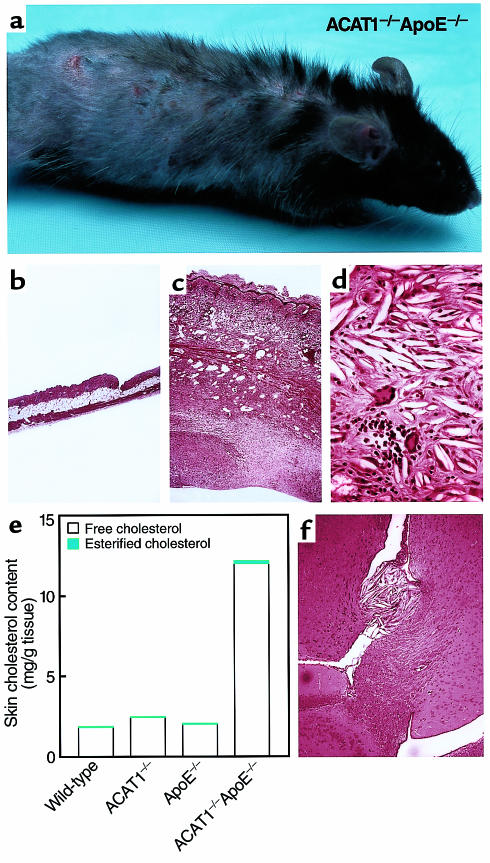
Figure 1.
Severe xanthomatosis in ACAT1–/–ApoE–/– mice. (a) Four-month-old ACAT1–/–ApoE–/– mouse fed a chow diet. Note the diffuse hair loss and excoriations due to pruritic skin lesions. (b–d) H&E-stained sections of skin from 4-month-old, chow-fed ACAT1+/+ApoE–/– (b, ×40) and ACAT1–/–ApoE–/– (c, ×40; d, ×100) mice. Marked thickening of skin was observed in ACAT1–/–ApoE–/– mice (c), with an intense inflammatory reaction disrupting the subcutaneous fat layer and muscularis carnosus. Severe fibrosis was present in the deeper layers of the skin. Higher magnification of the reticular dermis (d) revealed numerous cholesterol clefts and a pleomorphic inflammatory infiltrate that included multinucleated giant cells, neutrophils, mononuclear cells, and plasma cells. (e) Skin cholesterol content of 4-month-old, chow-fed mice (n = 2 in each group). (f) H&E-stained section of brain from ACAT1–/–ApoE–/– mouse fed a Western diet for 3 months. Note the free cholesterol crystal deposits and inflammatory infiltrate near the choroid plexus.

