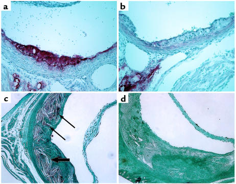
Figure 6.
Neutral lipid-poor atherosclerotic lesions in ACAT1-deficient mice. (a and b) Representative Oil-Red-O– stained aortic lesions from 6-month-old ACAT1+/+ApoE–/– (a) and ACAT1–/–ApoE–/– (b) mice fed a chow diet. Note the pale appearance of the ACAT1–/–ApoE–/– lesions due to a paucity of neutral lipids that stain with Oil Red O. (c and d) Representative sections of plastic-embedded, osmium tetroxide–stained aortic lesions from ACAT1+/+LDLR–/– (c) and ACAT1–/–LDLR–/– (d) mice fed an atherogenic diet for 2 months. Neutral lipid droplets (thin arrows) and numerous lucencies from cholesterol crystals (thick arrows) are apparent in the ACAT1+/+LDR–/– lesion (c). ACAT1–/–LDLR–/– lesions were devoid of osmium tetroxide staining and had fewer cholesterol crystals than ACAT1+/+LDLR–/– lesions.

