
Figure 3.
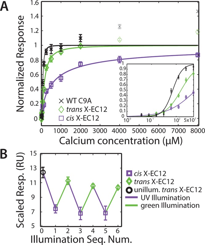
Characterization of photoswitch homodimeric binding. (A) Homodimeric binding monitored in SPR as a function of Ca2+ concentration. The data were fit to a Hill equation. Faded points contain significant nonspecific binding and were not used in the fits. Responses between flow cells were scaled to minimize a least-squares difference, and then mean values were normalized such that the fit value at [Ca2+] = ∞ was 1.0 (SI). Error bars are ±1 SD of the three active flow cells in the instrument after scaling and normalization. Inset shows fits at low Ca2+ concentrations. (B) Homodimeric binding monitored in SPR at 1 mM Ca2+, after repeated illumination cycles. Responses between flow cells were scaled to minimize a least-squares difference. Error bars are ±1 SD of the three active flow cells in the instrument after scaling.