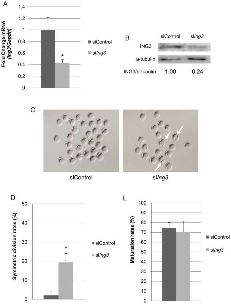
Figure 2. Effects of siIng3 injection on asymmetric cell division in mouse oocytes.
(A) Ing3 mRNA was significantly decreased in the fully grown GV oocytes injected with siIng3 (*p<0.05). (B) ING3 protein levels, after normalization to α-tubulin, were decreased in fully grown GV oocytes injected with siIng3. (C) Abnormal cell division was observed in several siIng3-injected oocytes at the MII stage (arrows). (D) In siIng3-injected oocytes, the rate of symmetric division was significantly increased as compared to that observed in siControl-injected oocytes (*p<0.05). (E) Maturation rates were not different between siIng3- and siControl-injected oocytes.

