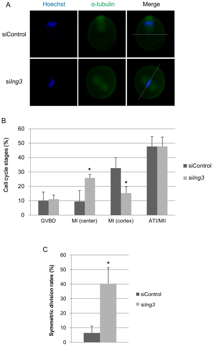
Figure 3. Effects of siIng3 injection on spindle migration in mouse oocytes.
(A) In siControl-injected oocytes, the spindle is located near the cortex at the MI stage after 9.5 h in culture. By contrast, in siIng3-injected oocytes, the spindle is located at the center of the oocyte at the MI stage after 9.5 h in culture. Bars indicate the central position of the oocyte. Green, α-tubulin; blue, chromatin. (B) In siControl-injected oocytes, the frequency of spindles located at the cortex was significantly increased. By contrast, in siIng3-injected oocytes, the frequency of spindles located at the center of the oocytes was significantly increased (*p<0.05). The oocytes that were extruding or had extruded a polar body were considered to be at the ATI or MII (ATI/MII) stage. (C) In siIng3-injected oocytes, the rate of symmetric division was significantly increased at the ATI/MII stages (*p<0.05).

