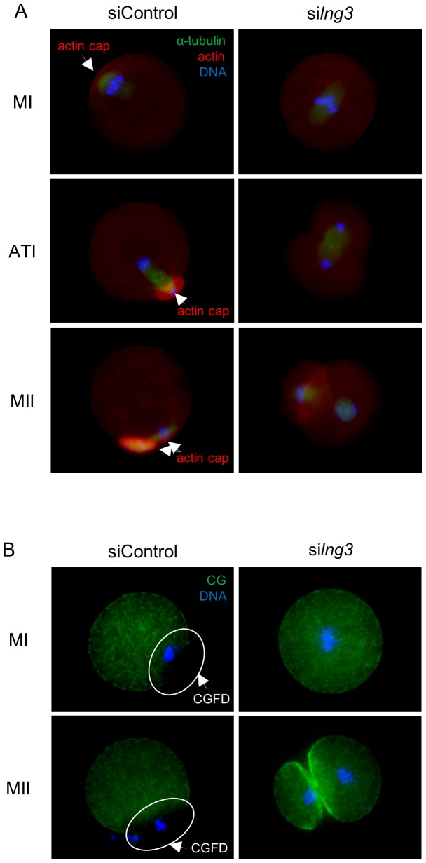
Figure 4. Effects of siIng3 injection on cortical reorganization in mouse oocytes.
(A) Actin cap formation was noted in siControl-injected oocytes at the MI, ATI, and MII stages. By contrast, no actin cap was formed in siIng3-injected oocytes at any stage. Arrows indicate the actin cap. Green, α-tubulin; red, actin; blue, chromatin. (B) CGs were absent in the cortex where the chromosomes were located at the MI and MII stages in siControl-injected oocytes. By contrast, in siIng3-injected oocytes, CGs were distributed throughout the cortex at the MI and MII stages, and were intensely localized in the region of cell adhesion at the MII stage. Circles and arrows denote the CGFD. Green, cortical granules; blue, chromatin.

