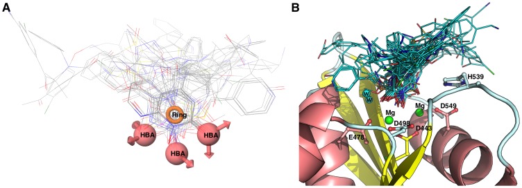
Figure 9. Comparison of the pharmacophore model with docking experiment.
Panel A. First ranked docking poses superimposed with the pharmacophore (Hypothesis 1, AAAR). Panel B. The first ranked docking pose is shown in the active site of RNase H and important residues are highlighted, including magnesium ions (green sphere) and water (cyan sphere).

