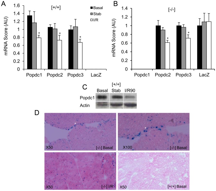
Figure 8. Popdc1 is down regulated by I/R.
(a) and (b) RT-qPCR of Popdc1-3 and LacZ mRNAs from hearts isolated upon heart removal (Basal), at the end of stabilization (Stab) and at 90 min reperfusion (I/R). N = 5/group. AU, arbitrary units, Mean ± SEM; *P<0.05 compared to Basal. (c) Western blots of Popdc1 in WT hearts (Popdc1, ∼68 kDa; Actin, ∼42 kDa). (d) Cytochemical staining of LacZ activity (blue nuclei). Note, a higher intensity in subendocardial cells (white asterisk). Eosin counterstaining illustrates the general morphology. Images were captured at X50 and X100 magnification, as specified.

