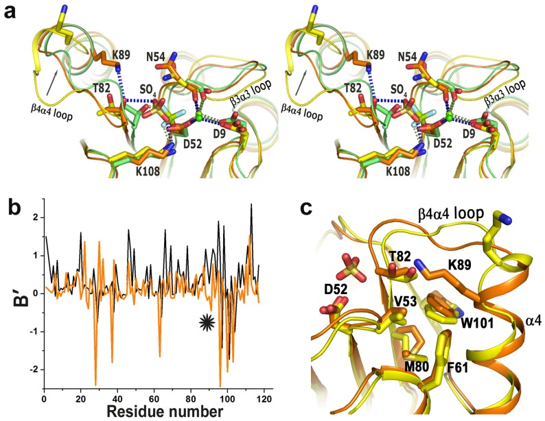
Figure 5. Structure of VcCheY4 in free and sulphated states.
(a) stereo view of the superposition of VcCheY4free (yellow) and VcCheY4sulf (orange) on activated VcCheY3-BeF3 − (green) showing the location and the interactions of the sulfate ion in VcCheY4sulf, relative movement of T82, hydrogen bond between K89 and T82 in VcCheY4sulf and the interactions of the metal ion with the neighbouring residues; (b) B′ plot of VcCheY4free (black) and VcCheY4sulf (orange) showing reduction of flexibility of the β4α4 loop (*) in VcCheY4sulf; (c) superposition of VcCheY4free (yellow) on VcCheY4sulf (orange) showing the conformational difference at the β4α4 loop and packing of W101 in its exclusive ‘in’ position.

