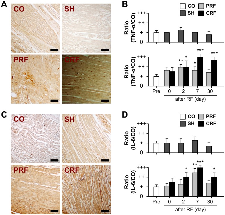
Figure 6. Up-regulation of TNF-α and IL-6 for RF-induced peripheral nerve injury.
RF, radiofrequency. The expression of TNF-α and IL-6 proteins in rat sciatic nerves with no treatment (CO), no current (SH), PRF and CRF was visualized by immunohistochemistry before stimulation (Pre) and on days 0, 2, 7 and 30 after stimulation. (A) Representative immunohistochemistry images on day 7 after RF treatment are shown. CO, control; SH, sham; PRF, pulsed RF; CRF, continuous RF. Scale bar = 100 µm. (B) The SH group showed no significant changes in TNF-α expression over the 30-day experimental period as compared to the CO group. In contrast, TNF-α expression was up-regulated immediately after RF treatment. This up-regulation persisted until days 7 and 30 after the PRF and CRF treatments, respectively. (C) Changes in IL-6 expression were similar to those observed for TNF-α. Scale bar = 100 µm. (D) IL-6 immunoreactivity reached a maximum on day 7 after exposure to PRF (P<0.005) and CRF (P<0.0001). Data shown are the mean and SD of six mice (n = 18). *P<0.05, **P<0.005, ***P<0.0001 vs. CO.

