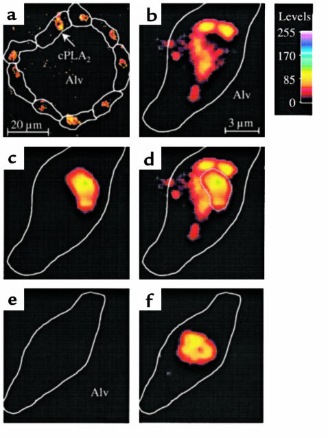
Figure 6.
Translocation of cPLA2 in single alveoli. An alveolus was infused with 1,000 U/mL TNF-α (a–d) or with Ringer’s solution (e and f). Then the alveolus was stained for both the immunofluorescence of cPLA2 (a, b, e) and the nuclear fluorescence of HOECHST 33324 (c and f). The alveolus was washed and imaged at low (a) and high (b–f) magnifications as indicated by the scale bars. Color code shows fluorescence intensities. Cell margins identified by bright-field microscopy are depicted by line sketches. Alv, alveolar lumen. Images for cPLA2 and nuclear fluorescence were combined by image overlay (d, f). Nuclear outline is indicated by dotted line (d). Replicated 4 times.

