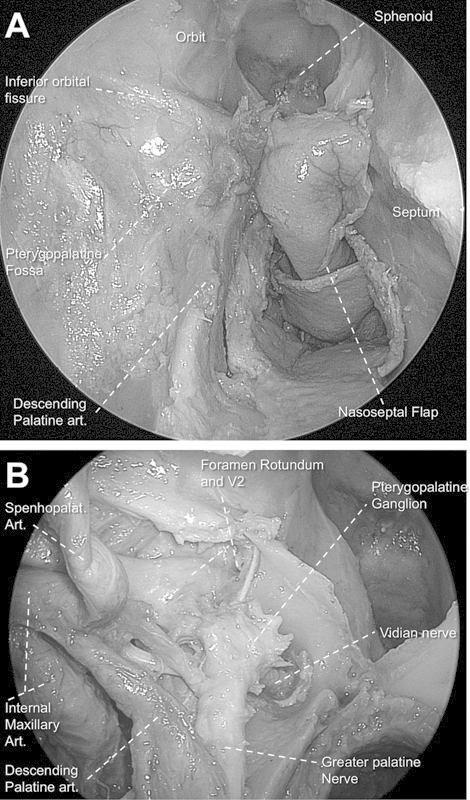
Fig. 1.

(A) Endoscopic view of right pterygopalatine fossa still covered by periosteum (0-degree endoscope). Note the descending palatine artery after removal of the surrounding bony sheath. (B) View after opening periosteum and removal of fat tissue of pterygopalatine fossa. Note the communication between vidian nerve, pterygoid ganglion, V2, and greater palatine nerve.
