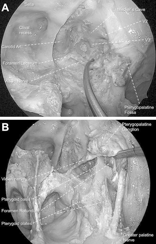
Fig. 2.

(A) Endoscopic transpterygoid approach to the Meckel cave in the left side (0-degree endoscope). Note that the vidian nerve is preserved. (B) View after unlocking the greater palatine nerve from its bony sheath. Note that the lateral mobilization of the neural structures of the pterygopalatine fossa allows an optimal exposure of all pterygoid base (partially drilled) and plates.
