Figure 1.
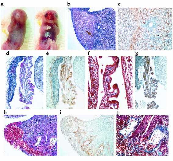
Analysis of an E17.5dpc FVII–/–/PC–/– embryo. (a) Gross appearance displaying significant internal and peripheral hemorrhage and edema. (b–c) H&E staining of liver sections indicating areas of fibrosis within 1 of the lobes (arrow) (b); anti-fibrinogen immunostaining of parallel liver sections demonstrating interstitial fibrin deposition (brown) around these fibrotic patches (c). (d–g) Histological analyses of hindbrain sections indicating areas of hemorrhage (d), fibrin deposition (e), collagen deposition (blue) (f), and the presence of CD45-positive leukocytes (brown) (g). (h–j) Histological analyses of atrial sections confirming the presence of blood in the atria (h), fibrin deposition (i), and internal collagen invasion around these areas of fibrin (j).

