
Fig. 1.
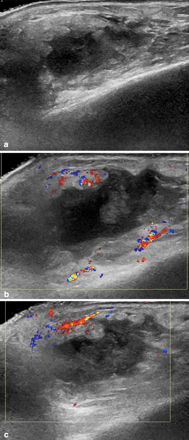
Parotid abscess. Ultrasonography shows a hypo-anechoic lesion, with irregular margins (a) and peripheral hypervascularity was detected on color Doppler examination (b, c)
Official websites use .gov
A
.gov website belongs to an official
government organization in the United States.
Secure .gov websites use HTTPS
A lock (
) or https:// means you've safely
connected to the .gov website. Share sensitive
information only on official, secure websites.
Parotid abscess. Ultrasonography shows a hypo-anechoic lesion, with irregular margins (a) and peripheral hypervascularity was detected on color Doppler examination (b, c)